The medial collateral ligament, or MCL, is one of the four major ligaments that stabilize the knee joint. MCL injuries are among the most frequent knee injuries in athletes and active adults, yet many people know very little about this critical structure. Understanding how the MCL works and what happens when it is damaged can help you protect your knees and recover more effectively.
1. It Is a Key Stabilizer on the Inner Knee
The medial collateral ligament runs along the inner side of your knee. It connects the bottom of the thighbone to the top of the shinbone. Its primary job is to resist forces that push the knee inward, a motion called valgus stress. Without a functioning MCL, the knee becomes unstable during side-to-side movements. This ligament works alongside the anterior cruciate ligament, posterior cruciate ligament, and lateral collateral ligament to keep the joint secure. Every time you plant your foot and change direction, your MCL helps absorb stress. Strengthening the muscles around the knee is one practical way to support this ligament during daily activities and sports.
2. MCL Injuries Are Graded on a Three-Level Scale
Doctors classify MCL injuries into three grades based on severity. A Grade I sprain involves mild stretching with microscopic tears but no significant laxity. Grade II means a partial tear with noticeable looseness when the knee is examined. Grade III is a complete tear where the ligament no longer provides stability. Research indicates that conservative treatment of Grade II sprains yields generally good long-term outcomes despite some residual laxity, while Grade III sprains often result in persistent instability and muscle weakness. Knowing your injury grade is essential because it directly shapes your treatment plan and expected recovery timeline.
3. Contact Sports Carry the Highest Risk
Sports that involve tackling, cutting, and sudden direction changes put the MCL under enormous stress. Football, soccer, hockey, and rugby are among the most common culprits. A direct blow to the outside of the knee forces it inward and can overload the ligament. Skiing is another well-known risk because falls can trap the leg in an awkward position. Non-contact injuries also occur during quick pivots when the foot is planted. Athletes in high-risk sports should consider wearing supportive braces and practicing proper landing mechanics to reduce their chances of an MCL tear.
4. Symptoms Often Appear Immediately After Injury
Most people feel a distinct pop or tearing sensation on the inner knee when the MCL is damaged. Swelling typically develops within a few hours and may be localized along the inner joint line. Pain is usually sharpest when pressing on the medial side or when the knee is stressed sideways. In more severe tears, the knee may feel wobbly or give way during walking. Some people also notice stiffness that makes bending the knee difficult. If you experience these symptoms after a knee injury, applying ice and seeking a medical evaluation promptly can help prevent further damage.
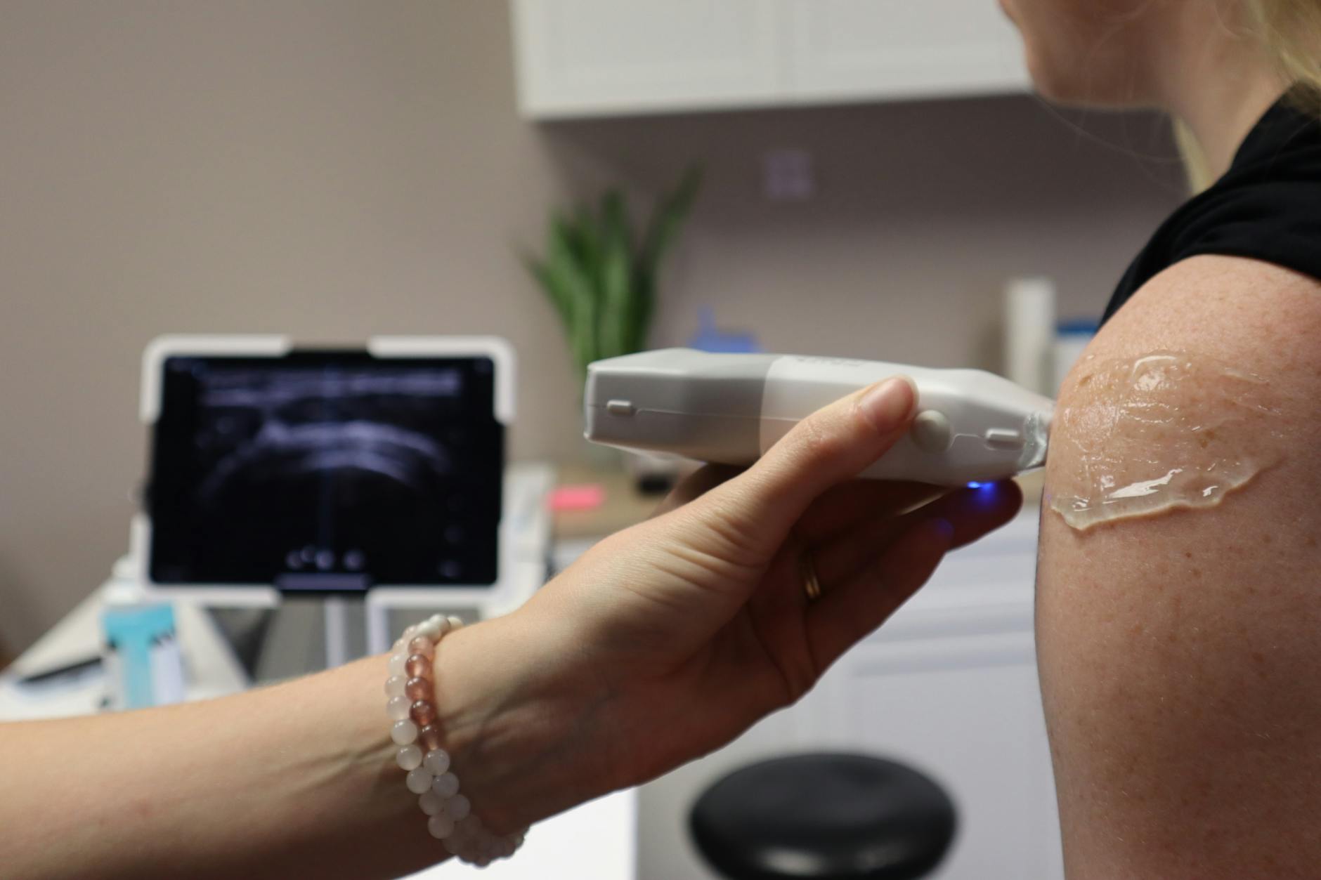
5. Diagnosis Usually Involves Physical Tests and Imaging
A healthcare provider can often diagnose an MCL injury through a hands-on examination. The valgus stress test, which applies gentle pressure to the outer knee while the leg is slightly bent, is the most common clinical assessment. If the knee opens up more than expected on the inner side, an MCL tear is likely. MRI scans are frequently ordered to confirm the diagnosis and check for damage to other structures. X-rays may be used to rule out fractures. Getting an accurate diagnosis early is important because it helps your doctor determine whether you need conservative care or a more advanced intervention.
6. Most Isolated MCL Injuries Heal Without Surgery
One encouraging fact about the medial collateral ligament is that it has a relatively good blood supply. This means it can often heal on its own with proper rest and rehabilitation. Grade I and most Grade II sprains are typically managed with bracing, physical therapy, and a gradual return to activity. Ice, compression, and elevation help control early swelling. Anti-inflammatory medications may ease pain during recovery. The healing timeline ranges from two to eight weeks depending on severity. Following your physical therapist’s exercise program closely is the single most important step for ensuring a strong recovery without lingering weakness.
7. Combined Injuries May Require Surgical Intervention
When an MCL tear occurs alongside damage to the ACL or meniscus, surgery becomes a more likely option. These multi-ligament injuries compromise overall knee stability in ways that conservative treatment alone cannot address. Studies suggest that MCL repair demonstrates higher rates of postoperative knee stiffness and failure compared to reconstruction, though both approaches improve patient-reported outcomes. Your surgeon will consider the specific combination of damaged structures, your activity level, and your long-term goals. If surgery is recommended, discuss all available techniques and their expected outcomes with your orthopedic specialist before making a decision.
8. Reconstruction Techniques Have Advanced Significantly
Surgical reconstruction of the MCL has evolved considerably over the past decade. Modern approaches focus on restoring the ligament’s natural anatomy as precisely as possible. Clinical evidence shows that double-bundle MCL reconstruction techniques using allografts significantly reduce medial joint opening and rotational instability in chronic cases. Some surgeons also use separate grafts to reconstruct the superficial MCL and the posterior oblique ligament individually. These anatomic methods aim to replicate the original ligament’s function rather than simply tightening the joint. If you are facing chronic MCL instability that has not responded to bracing and therapy, asking your surgeon about these newer reconstruction options is a worthwhile conversation.
9. Rehabilitation Follows a Structured Progression
Whether you are recovering from a mild sprain or a surgical reconstruction, rehab follows a phased approach. The early phase focuses on reducing swelling, restoring range of motion, and protecting the healing tissue. Gentle exercises like heel slides and straight-leg raises are common starting points. The middle phase introduces strengthening work for the quadriceps, hamstrings, and hip muscles. Balance and proprioception drills help retrain the knee’s sense of position. The final phase involves sport-specific movements like cutting, jumping, and agility drills. Rushing through these stages increases the risk of reinjury, so patience and consistency with your rehab program are essential for a lasting recovery.
10. Prevention Strategies Can Reduce Future Risk
Protecting the medial collateral ligament starts with building strong, balanced muscles around the knee. Exercises that target the quadriceps, hamstrings, and hip abductors help absorb forces before they reach the ligament. Neuromuscular training programs that emphasize proper landing and cutting mechanics have shown promise in reducing knee injury rates among athletes. Warming up thoroughly before activity increases blood flow and improves tissue flexibility. Wearing a hinged knee brace during high-risk sports may offer additional protection, especially if you have a history of MCL injuries. Making these habits part of your regular routine is one of the most effective ways to keep your knees healthy long-term.
The medial collateral ligament plays a vital role in knee stability, and understanding its function helps you make smarter decisions about prevention and treatment. Whether you are an athlete or simply someone who values mobility, talk to your doctor or physical therapist about a personalized plan to keep your knees strong and resilient.
Medical Disclaimer: This article is for informational purposes only and does not constitute medical advice. Always consult a qualified healthcare professional before making health decisions.


