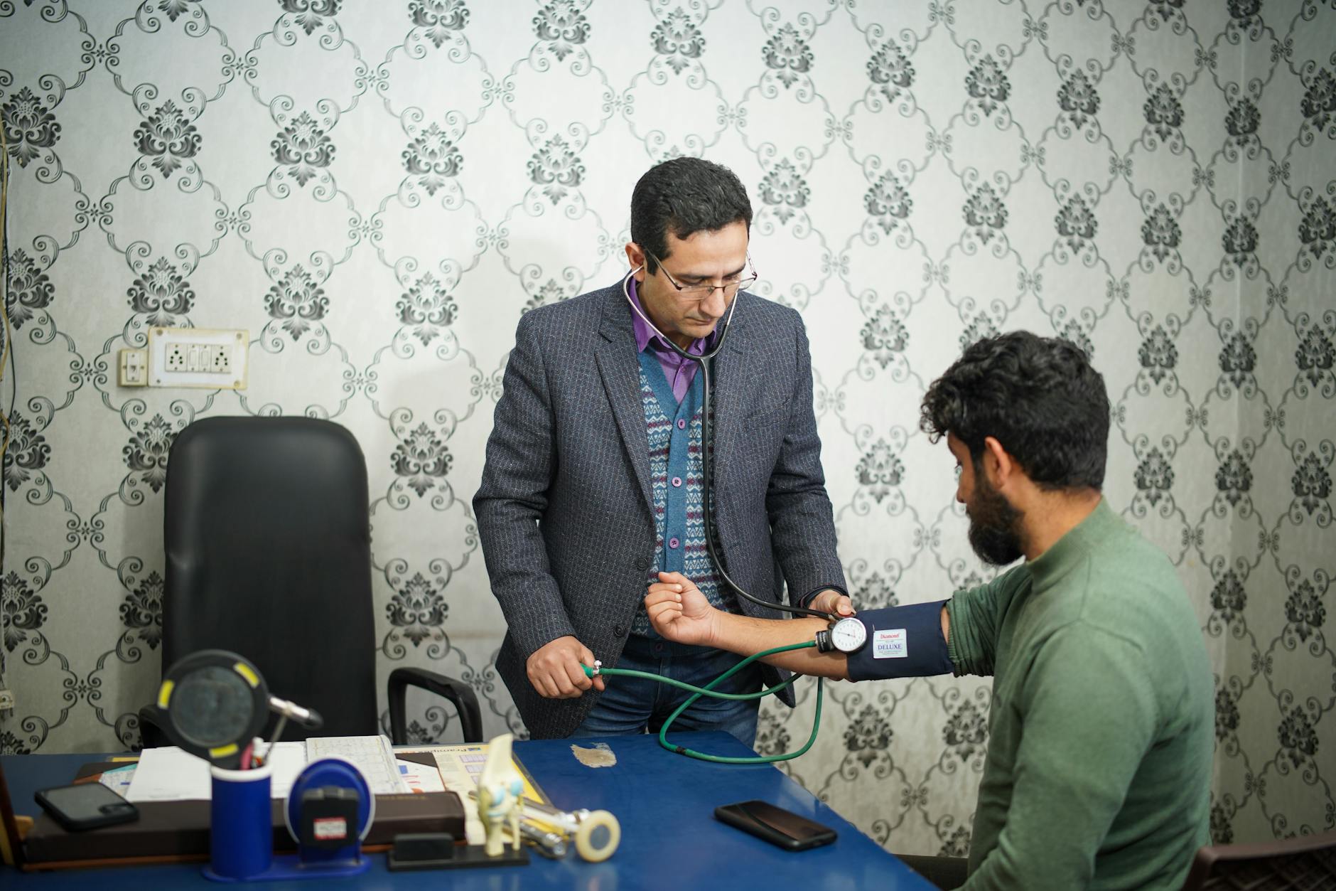
Patients walk into my urology clinic trembling because they googled the instrument we use to look inside their bladder. The metal tubes shown in vintage medical illustrations stopped being the standard decades ago. Today we use flexible optics that slide through the urethra with entirely tolerable friction.
1. The Anticipation Dwarfs the Reality
I hear the exact same phrase every Tuesday morning. “Doc, I haven’t slept since you told me what this was.” Most people construct an intricate torture chamber in their heads. But the transit time through the sphincter takes exactly four seconds. You feel a bizarre pressure. Then the lens clears the opening.
2. Blood in the Bowl Means We Look
General practitioners often write off a single episode of pink urine as a passing infection. They prescribe antibiotics and send you out the door. As a urologist, I witness the grim fallout of that delay. Microscopic blood demands a physical look inside the lining. A 2023 systematic review confirmed this optical inspection catches bladder cancer with up to 100 percent sensitivity, completely outperforming standard CT scans. We do not guess with hematuria. We illuminate the mucosa and trace every millimeter of the dome. Sometimes the tissue looks angry when it is just irritated.
3. The Numbing Agent Does Heavy Lifting
Most articles will tell you the procedure is painless. That framing misses the point entirely. You absolutely feel the intrusion. We push a generous volume of lidocaine jelly directly into the urethra before we even touch the scope. This viscous fluid serves two distinct purposes simultaneously. It freezes the nerve endings in the superficial mucosa. The gel also acts as a hydraulic wedge that gently forces the urethral walls apart so the lens glides rather than scrapes. Men tend to tense up right as the tip reaches the prostate. I tell them to drop their shoulders and wiggle their toes. Why do I ask for toe wiggling? It distracts the brain from pelvic floor guarding. The textbook describes a smooth insertion curve. The exam room reality involves steering through a tight, muscular bend that intensely resents being pushed open. I watched a guy grip the table edges until his knuckles turned white before the scope even touched him. He was bracing for absolute agony. Five minutes later he was watching the monitor with me, fascinated by the thick trabeculation of his own bladder wall. He asked if he could keep a printed picture of it.
The sting happens.
It just fades faster than your adrenaline does.
We pull the scope out slowly to check the sphincter function on the way out, and by the time you sit up, the sharpest sensation is already a memory.
4. Changing the Color of the Light Changes the Diagnosis
Standard white light washes out flat lesions. We miss things. A patient might come back six months later with a recurrent tumor we simply could not see against the pale pink background of the bladder wall. Now we inject a specialized dye an hour before the procedure. When we switch our light source to blue, malignant cells glow fluorescent pink. Clinical data from 2022 demonstrated this optical shift catches flat carcinoma in situ that white light completely obscures. You see the glowing margins immediately. It feels exactly like cheating.
5. The End of the Sterilization Conveyor Belt
We used to send every scope down to processing after a case. The turnaround took hours. Now I grab a sterile, single-use package off the shelf for almost every outpatient case. Recent hospital metrics proved these disposable optics dropped post-procedure complication rates by sixty percent. You are getting an instrument that has never touched another human being. The optics are incredibly sharp. The bending mechanism feels crisp and responsive in my hands. We toss the entire unit in the biohazard bin when we finish. No cross-contamination risk exists.
6. The Prostate Casts a Shadow
I can usually tell what I am going to find before the camera even clears the membranous urethra. The man shuffles into the room with a hesitant gait, sits down, and immediately crosses his legs. He looks thoroughly exhausted. I know his prostate is blocking the exit. The cystoscope just provides the geographic proof. We slide past the sphincter and suddenly hit a wall of obstructing adenoma kissing in the midline. The textbook calls this benign prostatic hyperplasia. I call it the reason a man wakes up four times a night to pee drops.
7. We Do Not Just Look Anymore
Historically, this was purely a scouting mission. We would look around, take some notes, and schedule you for a surgery two weeks later. That outdated model wastes everyone’s time. If I find a tight, scarred bladder neck causing your retention, we often fix it right then. We swap the diagnostic lens for a microscopic cutting tool. A 2023 urology trial evaluated this exact immediate-incision approach, finding men achieved massive improvements in urine flow without needing a second trip to the operating room. Men wake up from the mild sedation and instantly realize their bladder is empty. “It feels like a massive weight just fell out of my stomach,” one man told me last month. He had been unknowingly carrying a liter of residual urine for a year, destroying his kidneys in the process. We slit the dense scar tissue at the six o’clock and twelve o’clock positions. The neck springs open. The relief is mechanical and instant. Sometimes we do not fully understand why one man scars down after a mild infection while another heals perfectly. The fibrotic cascade in the pelvis remains stubbornly unpredictable to researchers. But the mechanical solution works beautifully.
8. Your First Void Will Burn
Nobody likes this part. The local anesthesia wears off exactly when your bladder demands to be emptied. The acidic urine hits the microscopic abrasions in your urethra. It feels like passing broken glass. This sharp sensation rarely lasts beyond the third trip to the toilet.
9. The Geography of Your Pelvis
Patients expect the inside of their bladder to look like a perfectly smooth balloon. It rarely does. When I inflate the organ with sterile water, the wall reveals thick, interlacing muscle bundles. (We call these trabeculations, a sign your bladder has been working too hard against out-flow resistance.) You can actually watch the tiny ureteral orifices squirting rhythmic jets of yellow urine from the kidneys into the clear irrigation fluid. It is a dynamic, constantly moving cavern, not a static storage tank.
10. The Monitor Tells No Lies
We do not wait for lab pathology to tell us if there is a massive tumor. The high-definition camera displays the reality on a screen two feet from your face. I watch my patients stare blankly at the monitor. Sometimes we see a delicate, frond-like growth swaying gently in the irrigation fluid. I must tell them right then that we are looking at cancer. There is nowhere to hide in that exam room. You walk out knowing exactly what we are up against.
The mechanics of this procedure are brutally straightforward, stripping away the mystery of your pelvic symptoms in minutes. Drink a gallon of water today to flush the irritated mucosa.
Medical Disclaimer: This article is for informational purposes only and does not constitute professional medical advice. Always consult a qualified healthcare professional before making changes to your health routine.


