A patient usually sits on the exam table for three weeks of failed antibiotic therapy before I see them. The sandfly that bit them in Costa Rica or Iraq is long dead, but the parasite it regurgitated into their bloodstream is quietly dissolving their tissue. Leishmaniasis behaves less like an infection and more like a slow-moving architectural collapse.
1. The eerie geometry of the painless crater
Most articles will tell you cutaneous leishmaniasis looks like a volcano-shaped sore. That framing misses the point entirely. Textbooks fixate on the geometry. But in the exam room, the defining trait is the eerie lack of pain. You stare down at a gaping wound on your leg that should have you screaming. Yet you barely notice it. “It just looks angry, doc, but it really doesn’t hurt at all,” a young botanist told me last year after returning from Peru. She had spent a month putting Neosporin on a parasite that was literally eating her macrophages. The raised, rolled border feels firm to the touch. Inside that border, the tissue resembles ground meat.
2. The antibacterial trap
Before patients end up in my infectious disease clinic, they usually spend a month on the wrong medication. Family doctors see a red, oozing bump and reflexively prescribe cephalexin. When that fails, they try doxycycline. I don’t blame them, because staph infections are common and sandfly parasites are not. We waste months treating a protozoan with antibacterial drugs that do absolutely nothing.
3. The ghost in the bone marrow
Cutaneous disease steals your skin, but visceral disease steals your marrow. Kala-azar, the old name for it, translates to black fever. I remember walking into room four to see a young man who had been traveling through Brazil. His chart listed unexplained fevers and unrelenting weight loss. Before the blood work even came back, I knew. His skin had a strange, hyper-pigmented gray duskiness to it. His abdomen looked slightly pregnant. The spleen had enlarged so massively it crossed the midline of his belly, pressing against his other organs. He was bleeding from his gums. The parasite had migrated from the initial bite site directly into his reticuloendothelial system, silently hijacking his liver. We eventually confirmed the diagnosis using rK39 rapid tests, which identify targeted antibodies from a simple blood draw. The physical presentation alone was unmistakable, though. Without treatment, visceral leishmaniasis kills almost everyone it touches. The parasites aggressively fill the bone marrow spaces until there is simply no physical room left to manufacture red blood cells or platelets. You bleed out. Or you catch a secondary bacterial infection your body is too utterly exhausted to fight. We admitted him immediately for intravenous therapy. It took weeks of intensive care to clear the parasite from his system. He survived, but his spleen will never fully shrink back to its original size.
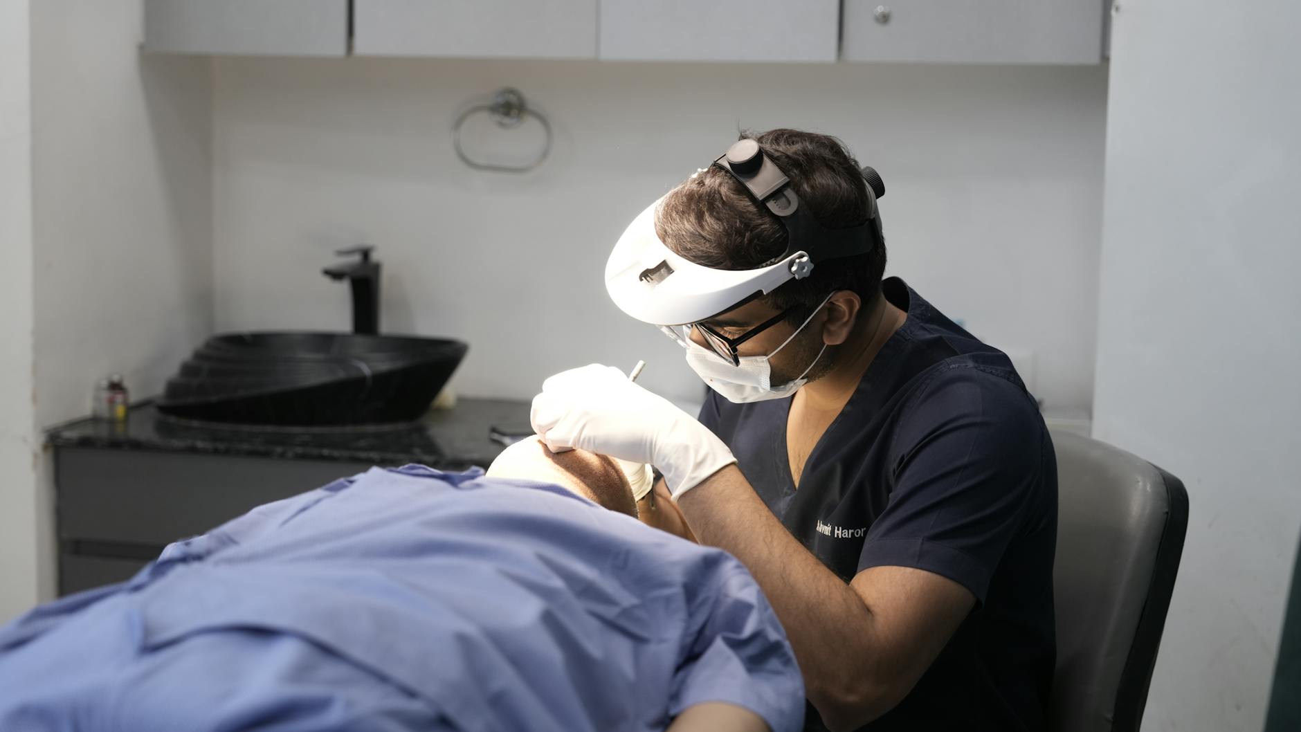
4. Scraping the living margins
How do we actually prove you have it? We take a scalpel and scrape the very inner edge of the ulcer. You cannot swab the pus in the middle. The parasites live in the healthy tissue at the margins, not the dead sludge in the center. We look under a microscope for amastigotes, which are little oval bodies hiding inside your white blood cells. Sometimes we find them immediately. Often, we have to send the tissue to a specialty lab for polymerase chain reaction testing. Getting a clean sample requires digging slightly deeper into the wound than the patient prefers. It hurts briefly, but it prevents months of misdiagnosis.
5. Liquid battery acid
We do not have gentle medicines for this disease. The oldest drugs we use contain heavy metals. We inject them directly into the vein, or worse, straight into the ulcer itself. “It felt like you shot battery acid into my leg,” a contractor who had been stationed in Afghanistan told me after his first round of sodium stibogluconate. We are slowly moving toward modern medications like liposomal amphotericin B. It works beautifully to clear the infection. But it requires daily intravenous infusions. It can also temporarily wreck your kidneys if we aren’t watching your creatinine levels closely.
6. A delayed collapse of the face
Healing the initial skin sore tricks you into feeling safe. Then, five years later, your nose starts bleeding. This is mucocutaneous leishmaniasis. The parasite goes dormant, travels through your lymphatic system, and wakes up inside the cartilage of your nasal septum or palate. It dissolves the structural framework of your face. (I once had a patient whose voice changed entirely because the roof of his mouth was quietly disintegrating.) The bizarre part is how selective the parasite behaves. Strains like Leishmania braziliensis possess a strange affinity for mucosal tissue, though we still don’t entirely understand the exact molecular trigger that wakes them up years after the initial bite. They just wait. And when they activate, they cause massive, destructive inflammation that melts cartilage into scar tissue. We often need to use DNA amplification techniques on the initial skin biopsy to figure out exactly which strain we are fighting. If you contracted a mucosal-associated variant in South America, we have to treat you aggressively with systemic drugs immediately. We cannot just let the skin ulcer heal on its own. The risk of facial disfigurement a decade later is simply too high to ignore. Even a tiny, innocent-looking bump on your arm requires full-force intravenous medication if the DNA test flags a dangerous strain. The cartilage in your nose has very poor blood supply, which makes it an ideal hiding spot for pathogens. By the time you notice a persistent stuffy nose or a minor nosebleed, the structural damage is already well underway. Reconstructive surgery is often the only option once the infection finally burns out.
7. Total silence at the ankle
Patients constantly tell me they never felt a bite. They expect an insect like a mosquito, which whines in your ear and leaves an itchy welt. Sandflies are a third the size of a mosquito. They make absolutely no noise. They fly low to the ground. Their bite doesn’t itch immediately. You never swat them away. They feed in total silence.
8. Melting geographic boundaries
Historically, doctors thought of this strictly as a tropical disease. That is no longer true. I am seeing cases acquired in Texas and Oklahoma from people who haven’t crossed an international border in a decade. The vector insects are expanding their geographic footprint as average temperatures warm. Dogs carry the parasite, too. An infected foxhound in the United States can be bitten by a local sandfly. That same fly then bites a human. The historical boundaries of infectious disease are melting right in front of us.
9. The cellular nursery
Your body tries desperately to wall off the parasite. It builds a granuloma, which is a dense cluster of immune cells meant to trap invaders.
But Leishmania actually prefers to live inside the very macrophages sent to destroy it.
It turns your primary defense mechanism into a nursery. The tissue damage you see on your skin isn’t mostly from the parasite eating you. It is your own immune system indiscriminately carpet-bombing the area trying to flush out an enemy that is hiding inside its own troops. The inflammation causes the surrounding skin to literally cave in on itself.
10. The permanent burn
Even after the parasite is eradicated, the physical evidence stays behind forever. The ulcers heal from the center outward. They leave a distinct, sunken, hairless scar that looks remarkably like a burn mark. It never repigments to match your surrounding skin. In places where the disease is endemic, these scars carry heavy social stigma. Here, they just serve as permanent, quiet reminders. You carry the mark of an insect that bit you on a Tuesday evening in a jungle thousands of miles away. The skin closes, but it never forgets the invasion.
The parasites that cause this disease operate on a timeline of months and years, easily outlasting basic clinical suspicion. Finding the correct diagnosis requires looking past the surface wound and interrogating the geographical history of the blood.
Medical Disclaimer: This article is for informational purposes only and does not constitute professional medical advice. Always consult a qualified healthcare professional before making changes to your health routine.


