Most patients assume their chest cavity holds two perfectly identical, hollow balloons. The physical reality of human breathing is a chaotic, asymmetrical plumbing system constantly fighting gravity and microscopic debris.
1. The Right Middle Lobe Problem
Textbooks map out three lobes on the right and two on the left. In the exam room, that falls apart. The right middle lobe sits like a tiny wedge, constantly battered by surrounding lymph nodes. When a patient coughs for six straight weeks, I listen there first. It acts like a trap.
2. The Apex Rises Higher Than You Think
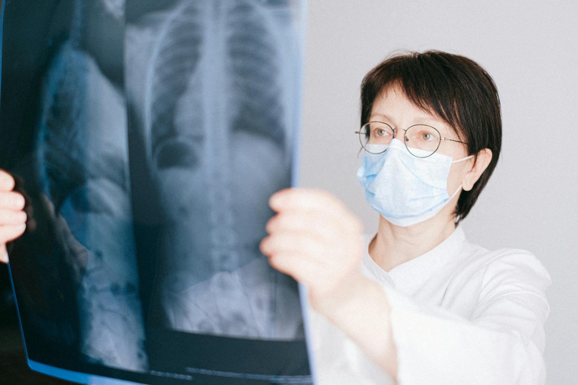
“My shoulder blade aches every time I take a deep breath.” A patient said this to me yesterday morning. She figured it was a torn muscle from hauling yard waste. The actual anatomy behaves entirely differently. Your lung apex extends well above the collarbone, reaching up into the soft base of your neck. General practitioners routinely miss early masses in this upper region. Standard plain chest films simply can’t see past the dense clavicle bone to spot small nodules hiding behind the joint. Patients spend months trying physical therapy for a presumed shoulder injury before anyone orders advanced imaging. We find the true cause later. A thoracic specialist always checks the apices carefully on a high-resolution CT scan.
3. The Fissures That Rarely Exist
Anatomy diagrams draw thick black lines separating the lobes. We call these fissures. They are supposed to let the segments slide against each other smoothly, like wet glass. In a living adult, those neat divisions rarely exist. I see fused tissues in at least half the bronchoscopies I perform weekly. Decades of minor viral infections, inhaled city dust, and normal biological aging act like glue. They weld the barriers together. This completely alters how pneumonia spreads through the chest cavity. If a fissure is incomplete, infected fluid simply leaks from the upper lobe directly down into the lower one. Most articles will tell you bacterial infections stay safely confined to one region. That framing misses the point. The internal boundaries are highly porous. We do not fully understand why some people maintain pristine anatomical separation while others fuse completely by their late thirties. Standard anatomical texts state the right lung possesses three distinct lobes separated by strict oblique and horizontal fissures. The reality on a surgeon’s table usually looks like a single blurred gradient of pinkish-gray mass. Inflammation ignores the map entirely. When a radiologist reads the scan, they have to guess where the upper lobe ends. The textbook neatness is a fiction.
4. The Left Side Surrenders Volume
Your left lung surrendered a massive chunk of real estate to accommodate the heart. We call this the cardiac notch. It leaves the lower lobe wedged awkwardly behind the pumping ventricle. Looking at advanced imaging models built from human CT data, the asymmetry jumps out at you. The left side is ten percent smaller. And that missing volume matters when you catch a bad respiratory virus. Reserve capacity drops. You feel short of breath much faster if the left side takes a direct hit. The right side simply has to work harder. I constantly see patients who tolerate right-sided pneumonia decently well but end up hospitalized when the exact same infection strikes the left lung.
5. The Pleural Vacuum
Your lungs are basically stuck to the inside of your rib cage by negative pressure. There is no physical attachment holding them open. A microscopic layer of fluid acts like water between two panes of glass. They slide, but they can’t pull apart. If a broken rib lets outside air into that space, the vacuum breaks. The lung collapses instantly.
6. The Tree Inside Your Chest
We talk about the bronchial tree because the internal branching pattern mimics a winter oak. The main trachea splits. Then it divides into lobar tubes. After that, dozens of narrow segmental pathways emerge. By the time oxygen reaches the alveoli, the vessels are microscopic. I remember examining a young man who worked the night shift in a commercial bakery. He said, “I just feel heavy right in the center of my breastbone.” I listened to his back. There was a faint, high-pitched whistle isolated entirely to the right lower lobe before we even ordered the imaging. A chest film later confirmed focal collapse. Inhaled flour dust had completely blocked a tertiary airway. The neural control regulating local ventilation, mapped out in historical anatomical reviews of lung parenchyma, was desperately trying to trigger a cough to clear it. But the angle of the right main bronchus is much steeper than the left.
Gravity usually wins.
Debris falls into the right side easily. A swallowed peanut almost always lands down in the right lower lobe. Dentists occasionally drop crowns down that exact same anatomical slide. We pull them out with tiny biopsy forceps. It’s fundamentally a plumbing issue dictated by the steep angle of the right pipe.
7. The Double Blood Supply
Most organs draw blood from a single vascular source. The lungs get two entirely separate deliveries. The massive pulmonary arteries carry depleted blood from the body to grab fresh oxygen. But the lung tissue itself needs constant cellular nourishment to survive. That fuel comes from the tiny bronchial arteries branching straight off the aorta. It functions as a brilliant biological backup system. If a massive clot blocks the main pulmonary artery, the organ usually survives the initial insult. Why? Because the secondary bronchial supply keeps the actual cellular structure alive until we can medically dissolve the blockage. You might feel terribly short of breath, but the underlying tissue holds on. It buys us time in the emergency room.
8. Cilia Act As An Escalator
You produce roughly a quart of mucus every single day. Millions of microscopic hairs line the major airways. They beat in a synchronized rhythm. This motion pushes a continuous sheet of sticky fluid up toward your vocal cords. You swallow it constantly. When someone smokes, those delicate hairs become chemically paralyzed. The escalator stops. Thick mucus simply pools in the lower dependent lobes. That stagnant pond turns into a massive breeding ground for aggressive bacteria. This failure is why smokers wake up hacking. They have to manually force the fluid upward because the tiny brooms quit working hours ago. The anatomy hasn’t changed, but the mechanical function is entirely broken.
9. The Tennis Court In Your Chest
If you flattened out every tiny air sac in an adult chest, it would cover half a tennis court. That staggering surface area allows oxygen to transfer into your blood fast enough to keep you conscious. We are born with only a tiny fraction of these sacs. They multiply rapidly during early childhood. Sometimes I catch myself wondering if damaged adult tissue could ever regenerate that way. (We see mild physiological improvements in patients who quit smoking, though true new cellular growth remains unproven.) Once the delicate walls between alveoli break down from emphysema, they form large useless pockets. Air gets trapped. You can breathe in, but you can’t exhale fully. The total surface area shrinks, leaving you starved for oxygen despite taking massive gasping breaths.
10. The Diaphragm Does The Heavy Lifting
We naturally fixate on the lungs themselves. But these are entirely passive organs. They possess zero muscle tissue. The diaphragm is a massive parachute-shaped dome sitting just beneath them. When it contracts, the thick central tendon pulls downward toward your stomach. This drops the pressure inside your chest cavity, physically sucking outside air through your nose. Patients with severe neuromuscular diseases rarely lose functional lung tissue. They just lose the sheer physical strength to pull the diaphragm downward. Without that underlying muscle dropping the floor of the chest, the healthy lungs above it stay completely deflated. The bellows can’t open. Breathing stops not because the lung fails, but because the engine driving it simply shuts off.
Your respiratory architecture dictates exactly how infections and inhaled particles settle in your chest. If you develop a persistent cough localized to one side of your back, ask your physician to listen to the dependent lobes.
Medical Disclaimer: This article is for informational purposes only and does not constitute professional medical advice. Always consult a qualified healthcare professional before making changes to your health routine.


