I saw three patients last summer who had been sent home from urgent care with a diagnosis of ringworm. They all had early-stage Lyme.
1. The textbook lie
Medical students are shown a perfect target lesion in their first dermatology rotation. A bright red outer ring, a crisp zone of clearing, and a dark red center. That textbook presentation is rarely what I actually see in the exam room. Most of the time it looks like a uniformly solid, slightly raised red blotch that someone assumes is a severe mosquito bite or a mild allergic reaction to a weed. This is exactly what gets missed at the GP level versus the specialist level. A rushed family doctor glances at a solid red patch on a thigh, prescribes a topical steroid cream, and moves on to the next room. By the time they end up in my office weeks later, the deep bone fatigue has already set in. I look closely at the spreading margins under the harsh clinic lights. Does the edge feel slightly warmer to the touch than the surrounding skin? Yes. Is it expanding outward by a few millimeters a day? Yes. That is erythema migrans. Waiting around for a perfect bullseye to form before starting antibiotics is a fast track to joint pain and neurological symptoms down the line. We treat what we see. And we see solid red flesh more often than we see neat concentric circles.
2. Fading doesn’t mean healing
The rash vanishes on its own. It happens without antibiotics, without creams, without any intervention at all. Patients think they’re cured. They aren’t. The spirochete bacteria simply moved from the dermal layers into the bloodstream. A disappearing red mark just means the localized skin reaction is over. The systemic infection is just getting started.
3. The purple bruise imposter
Sometimes the skin reacts with deep vascular congestion instead of bright surface inflammation. You might look down at your calf and see what looks like a spreading, violaceous bruise. Bhattarai et al. (2024) detailed a 69-year-old female who presented with a dark, purplish Lyme disease rash that completely lacked an outer ring. Her serology confirmed the infection, and doxycycline cleared it rapidly. I’ve had patients swear they just bumped their leg on a coffee table. But bruises change color as they heal. They go green, then muddy yellow. Erythema migrans doesn’t do that. It just expands outward, keeping that deep plum color active at the margins.
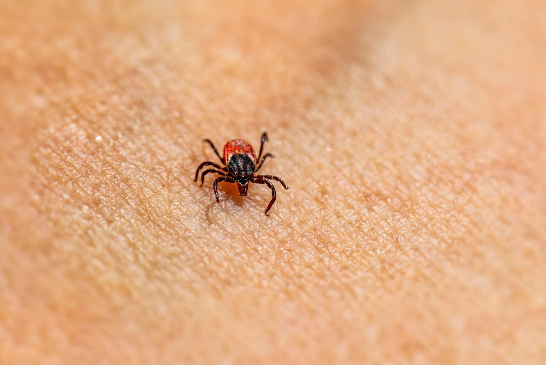
4. The spider bite illusion
The number of times I hear about imaginary arachnids is staggering. “I know it’s a spider bite because I felt a sharp pinch when I was gardening,” a woman told me last Tuesday. Spiders rarely bite humans unless physically trapped against the skin. Ticks, on the other hand, inject a local anesthetic so you don’t feel them burrow in. By the time the red lesion swells and becomes itchy, the tick has often fed and dropped off. The patient is left staring at a hot, red welt with a tiny central crust, fully convinced a brown recluse is living in their tomato plants.
5. Hiding in the hairline
Ticks crawl upward. They look for warm, moist, hidden places to feed quietly. You might be checking your arms and calves, but the rash is silently blooming behind your ear or buried under the hair at the nape of your neck. You usually only find it because your cervical lymph nodes swell up first.
6. Blisters at the center
Most articles will tell you erythema migrans is perfectly flat and smooth. That framing misses the point. The skin can absolutely blister. The intense local immune response to tick saliva and bacterial proteins occasionally causes tiny fluid-filled vesicles to form right in the middle of the red patch. Schünemann et al. (2013) recorded how atypical lesions present as vesicles, papules, or even lymphangitic streaks tracking up a limb. When those blisters pop, they crust over. It looks exactly like a staph infection, tricking everyone into treating the wrong bug.
7. The geographic mismatch
You would think people living in tick-heavy areas know what to look for.
They don’t. According to Ahern et al. (2012), while about 72 percent of people recognize a classic target lesion, they fail completely at identifying atypical blue-purple lesions or vesiculopustular forms. The public expects a neat, orderly circle on their skin. Biology is rarely neat.
8. The delayed arrival
A guy walked into my clinic complaining of summer flu. Sweats, aching knees, a stiff neck. He had a faint, oval-shaped pink smudge on his shoulder blade. I looked at that smudge and knew exactly what it was before the blood work even came back. Why? Because the rash doesn’t always show up the day you are bitten. It takes anywhere from three to thirty days for the bacteria to replicate enough in the dermal layers to trigger that visible red ring. He had been camping a month prior. The timeline fit perfectly, even though he hadn’t been in the woods for weeks.
9. Disseminated spots
What happens when the bacteria enter the bloodstream? You get multiple rashes. They pop up on your back, your stomach, your thighs. They are usually smaller than the original bite site and rarely have a central puncture mark. “It’s like somebody took a pink bingo stamper to my chest,” a young landscaper said to me last fall. These disseminated lesions mean the infection is no longer localized in one patch of skin. It is entirely systemic.
10. The lingering shadow
You take the standard course of doxycycline. You swallow the last pill. The systemic symptoms fade out, the brain fog lifts, but you look down at your calf and a faint, brownish shadow is still sitting right where the redness used to be. Patients panic when they see this. They assume the bacteria are still alive and actively eating through their connective tissues. The truth is much more mundane, though the exact mechanism of post-inflammatory hyperpigmentation in spirochetal infections is not fully understood yet. Your immune system carpet-bombed that localized area of skin to fight the invading Borrelia bacteria. (Collateral damage is inevitable when white blood cells degranulate in tight dermal spaces). The active infection is completely gone, eradicated by the antibiotics, but the heavy melanin deposits left behind from the inflammatory chaos remain lodged deep in the tissue. It can take months for your body’s macrophages to slowly clear out that excess pigment. The shadow is just a localized scar. It operates as a stubborn footprint of where a microscopic battle happened weeks ago. You don’t need a second round of harsh antibiotics for a footprint. You just wait for the skin cells to turn over and carry the debris away.
Medical Disclaimer: This article is for informational purposes only and does not constitute professional medical advice. Always consult a qualified healthcare professional before making changes to your health routine.


